Wechat QR code

TEL:400-654-1200

TEL:400-654-1200
Electroencephalography is a pattern obtained by magnifying and recording the spontaneous biological potential of the brain from the scalp through an instrument.
Normal value
Normal EEG can be divided into the following four types: α-shaped EEG, β-shaped EEG, low-voltage EEG, irregular EEG. EEG normal frequency and amplitude according to different, can be divided into two kinds of α-wave and β-wave.
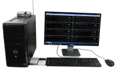
EEG machine
Clinical significance

Abnormal results: abnormal EEG can be divided into mild, moderate and severe abnormalities. (1) mild abnormal EEG α rhythm is very irregular or very unstable, eyes open suppression reaction disappeared or not significant. The amount of high-frequency β area or area appear. Q wave activity increased, Q activity in some parts of the dominant, and sometimes Q wave in all districts. Excessive ventilation after a high amplitude Q wave. (2) moderate abnormal EEG α section activity frequency slow down disappeared, there is obvious asymmetry. Diffuse Q activity dominant. Occurred paroxysmal Q wave activity. After hyperventilation, high-amplitude δ waves appear in groups or in groups. (3) Severe abnormal EEG diffuse Q and δ activity predominance, high voltage δ activity in the slow wave. Alpha rhythm disappears or slows down. Occurrence of paroxysmal δ wave. Spontaneous or induced high-amplitude spikes, sharp waves or spike slow synthesis wave. An outbreak of inhibitory activity or flat activity. EEG abnormalities on the diagnosis of the following diseases have some help: (1) disturbance of consciousness (drowsiness, coma, etc.). (2) intracranial space-occupying lesions: including brain tumors, brain abscesses, brain metastases and chronic subdural hematoma. (3) epilepsy. (4) traumatic brain injury: concussion, brain contusion and so on. (5) cerebrovascular disease: cerebral hemorrhage, cerebral thrombosis. (6) Intracranial inflammation and encephalopathy: viral encephalitis. Need to check the crowd: Suspected brain abnormalities in patients.